
Oct 1st – Walking the tight-rope between Bleeding and Clotting
On Oct 1, 2013, the Life Sciences Institute (LSI) at the University of British Columbia hosted the 13th LSI Café Scientifique. Over 60 interested community members, students and faculty gathered for an informal and participatory dialogue with LSI experts for new series of Cafes focused on Disease. The topic of this session was “Walking the tight-rope between Bleeding and Clotting”.
The Café featured members of the Centre for Blood Research, Dr. Ed Pryzdial, Scientist and Clinical Professor, department of Pathology and Laboratory Medicine and Dr. Shannon Jackson, Clinical Assistant Professor and Staff Hematologist, Providence Health Care
After injury to a blood vessel, your body has an intricate method to seal just the site of the leaky vessel by making a blood clot. Eventually the clot is dissolved to restore normal blood flow, so healing can occur. Thrombosis is the highly prevalent disease that results in blockage of a blood vessel when the precise balance between clot-forming and clot-dissolving tips to the former, resulting in a heart attack, deep vein thrombosis or stroke. On the other side of the balance, if not enough clot forms, serious bleeding may occur, such as in hemophilia. Drs. Jackson and Pryzdial tag-teamed to explain the blood clotting “tight-rope”, the treatment of perplexing patients dangling on the tight-rope and the development of a new clot-busting medicine.
To view the video link of the session, please click HERE.
To view more pictures from the event, please visit our Facebook page.
Seeing is Believing Series – Super-resolution Microscopy: Breaking the Diffraction Barrier
 On April 30, 2013, the Life Sciences Institute (LSI) at the University of British Columbia hosted the 12th LSI Café Scientifique. Over 60 interested community members, students and faculty gathered for an informal and participatory dialogue with LSI experts for the fifth session in the “Seeing Is Believing” series. The topic of the session was “Super-Resolution Microscopy: Breaking the Diffraction Barrier”.
The Café featured Dr. Ivan Robert Nabi, member of the Cell & Developmental Biology (CELL) Research Group and Department of Cellular & Physiological Sciences and Dr. Keng-Chang Chou from the Department of Chemistry.
Since the invention of compound microscope in 1590 by Zaccharias Janssen and his son Hans, microscopy has made great  contributions to the advancement of science. In the past 50 years, scientists have used a technique called “fluorescence microscopy” to observe the inner working of cells. In this technique, a light beam illuminates proteins marked with a fluorescent tag. By providing a glimpse of what goes wrong inside cells affected by disease, this technology has driven many important advances in biomedical research. Conventional fluorescence microscopy reveals many cellular features, but the tiniest structures – those that allow cells to communicate with each other and the outside environment – have been hidden in the haze caused by the diffraction of light (i.e. the diffraction barrier). The effect of light diffraction limits the resolution of an optical microscope to approximately half of the wavelength of light used. With the best optics, the resolution of fluorescence microscopy is limited to ~ 200 nanometres (nm), which cannot resolve many fine cellular structures.
Now, a revolutionary breakthrough has created a new type of microscope that cuts through this haze, breaking the diffraction barrier and bringing the tiniest cellular structures into sharp focus. Called “super-resolution” microscopy, this game-changing technology allows researchers to observe structures as small as 20 nm – just ten times the size of the largest proteins – and track them over time within a living cell. Stimulated emission depletion (STED) imaging uses a second doughnut-shaped laser beam to shrink the effective size of the imaging laser, lighting up a smaller region of fluorescent proteins. This provides outstanding lateral resolution of 50-70 nm, and rapid imaging in three dimensions without need for complex mathematical interpretation of the data. Localization microscopy approaches are based on the repeated activation of small numbers of discrete fluorophores whose precise localization is determined using a Gaussian fit of the point-spread function (PSF). Repeated activation of samples generates images whose X-Y resolution is on the order of 20 nm.
Super-resolution microscopy represents the next frontier of optical imaging for biological and health research applications. Using CFI-funded infrastructure, the imaging community at UBC will now develop a Super Resolution Core imaging unit to apply live cell super-resolution imaging to disease models.
A mounted image entitled “Caveolae at super-resolution”, supplied by Dr. Nabi, was given away as the door prize to a member of the audience.
The LSI Café Scientifique is co-sponsored by the Life Sciences Institute (LSI), Michael Smith Foundation for Health Research (MSFHR), Faculty of Medicine Research Office, Faculty of Science Dean’s Office, the Leica Corporation, the microscope company Systems for Research and Café Perugia (UBC Food Services).
The next Café Scientific that will continue the “Seeing is Believing” series will take place in fall of 2013.
To look at more pictures, please visit our facebook page.
To view the tape recording, please visit our Youtube channel.
Seeing is Believing Part 4, “Viewing the Biological World with X-rays and Magnetic Fields”
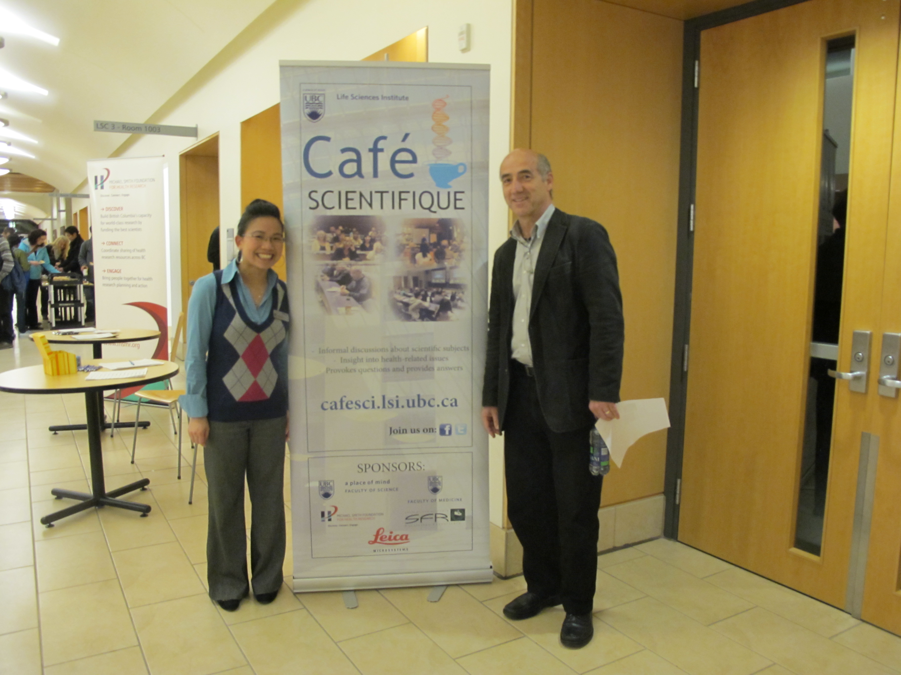 On Nov 14, 2012, the Life Sciences Institute (LSI) at the University of British Columbia hosted the 11th LSI Café Scientifique. Over 70 interested community members, students and faculty gathered for an informal and participatory dialogue with LSI experts for the fourth session in the “Seeing Is Believing” series. The topic of the session was “Viewing the Biological World with X-rays and Magnetic Fields”.
The Café featured Dr. Michael Murphy, a member of the Bacterial Adaptation & Response Networks (BARN) LSI Research Group and the Dept of Microbiology & Immunology, and Dr. Lawrence McIntosh, a member of the Chemical Biology of Disease (CBD) LSI Research Group and the Depts of Biochemistry & Molecular Biology and Chemistry.
Dr. Michael Murphy started the discussion explaining the ranges of sizes of objects, starting with a grain of rice and moving progressively smaller and smaller to show where biomolecules fit into the sequence of sizes. He then went on to show that X-ray crystallography provides a detailed atomic view of biological molecules. This visualization technique relies on producing small crystals of the biomolecule that are subject to an intense highly focused X-ray beam. The data collected from the X-ray beam interacting with the crystal is used to create three-dimensional maps of the electron density that describe the structure of the molecules. The resulting image is a snapshot of an average over all the molecules in the crystal. Multiple snapshots of the crystallized biomolecules in different states such as a free receptor versus the ligand bound form can be used to describe function of the proteins at a molecular level. Examples were taken from one of the systems studied in the Murphy lab, the human bacterial pathogen, multiple drug resistant Staphylococcus aureus. Bacteria need to acquire iron to grow but our bodies attempt to sequester iron to limit bacterial growth. Effective pathogens like S. aureus have developed specialized scavenger proteins to overcome this nutritional need by pirating iron from host proteins. These bacterial scavenger proteins are potential targets for the development of new antibacterial therapeutics and diagnostic drugs. Throughout the talk, computer generated protein structures were shown that highlighted the various regions of proteins using a variety of views. The talk ended with a brief question and answer session.
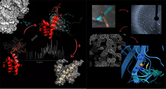 Dr. Lawrence McIntosh went on to explain that Nuclear Magnetic Resonance (NMR) spectroscopy is a “molecular microscope” that enables researchers to study the three-dimensional structures of proteins and thereby gain insights into their biological functions. NMR relies on measuring the energy required to “flip” the spins of nuclei aligned within a powerful magnet field. This energy depends on the chemical environment of the nucleus, and hence on the structure of protein. Importantly, proteins do not have static structures, as shown in the field by beautiful diagrams of ribbons and coils, but rather are highly flexible and dynamic. This flexibility, which can also be measured with NMR, is important for protein function. One such function is the self-inhibition of a transcription factor that balances the energetic cost of unfolding a helix structure of this protein with the benefit of binding DNA. This balance can be changed in response to cellular signals in order to turn genes on or off, working as a molecular switch. Dr. McIntosh’s talk also highlighted computer generated molecular models that showed the dynamic structure of the proteins he studies. At the end, he also answered questions from the audience.
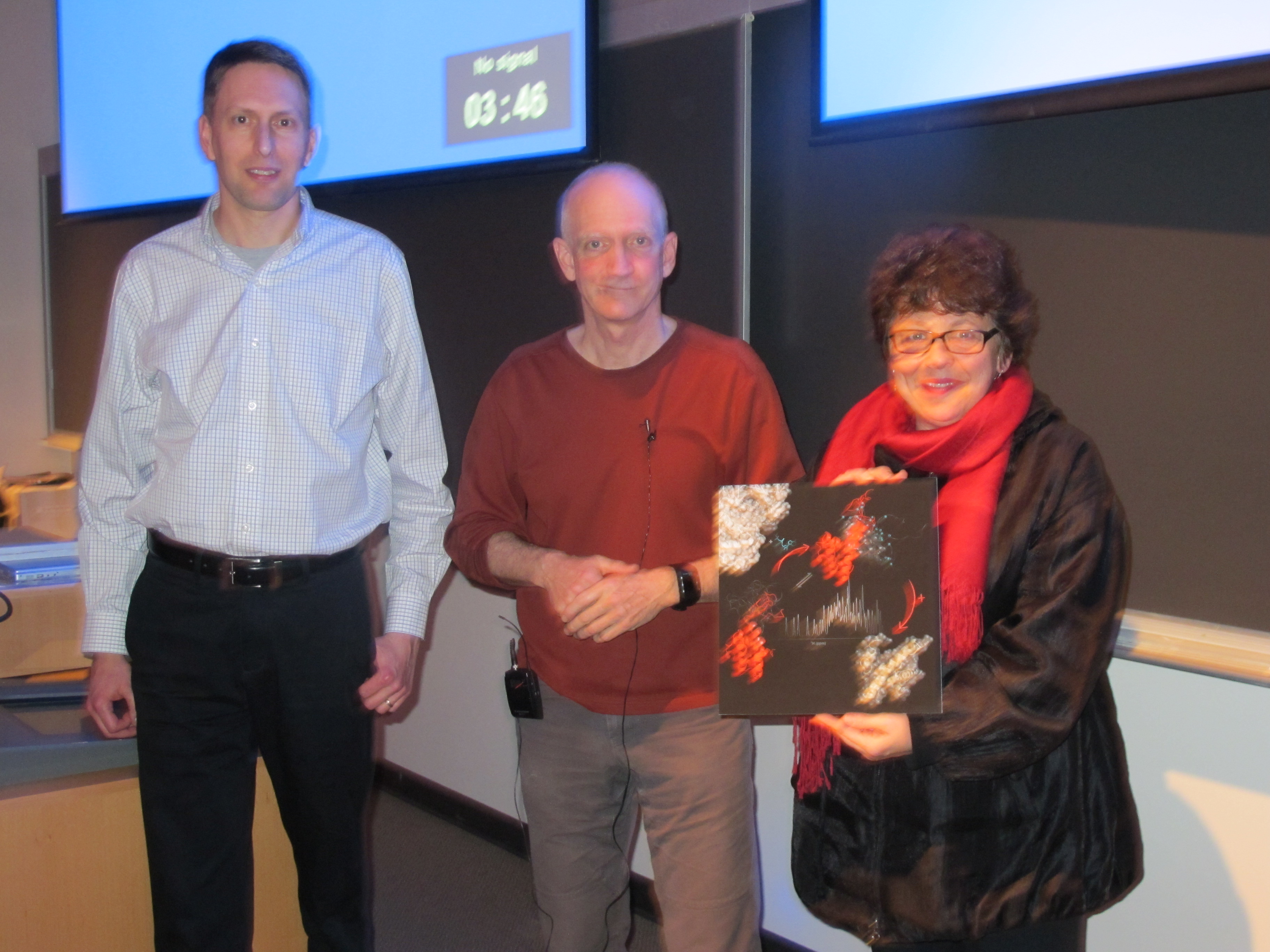 Both the Murphy lab and the McIntosh lab are aided by infrastructure (equipment) purchased by the Canada Foundation for Innovation grant to the ASTRID (“Advanced Structural Analysis of Re-emerging Infectious Diseases”) initiative, located in the Life Science Centre.
At the end of the talks, a mounted image entitled “Biological NMR Spectroscopy”, supplied by Dr. Lawrence McIntosh, was given away as the door prize to a member of the audience.
The LSI Café Scientifique is co-sponsored by the Life Sciences Institute (LSI), Michael Smith Foundation for Health Research (MSFHR), Faculty of Medicine Research Office, Faculty of Science Dean’s Office, the Leica Corporation, the microscope company Systems for Research and Café Perugia (UBC Food Services).
To look at more pictures, please visit our facebook page.
The next Café Scientific that will continue the “Seeing is Believing” series will take place in early 2013.
“Viewing the Biological World with X-rays and Magnetic Fields” by Dr. Michael Murphy
“Viewing the Biological World with X-rays and Magnetic Fields” by Dr. Lawrence McIntosh
Seeing Is Believing Part 3, “NanoSpace Invaders: Seeing into the subcellular world”
 On May 29, 2012, the Life Sciences Institute (LSI) at the University of British Columbia hosted the 10th LSI Café Scientifique. Over 50 interested community members, students and faculty gathered for an informal and participatory dialogue with LSI experts for the third session in the “Seeing Is Believing” series. The topic of the session was “NanoSpace Invaders: Seeing into the Subcellular World”.
The Café featured members of the Cell and Developmental Biology Research Group and the Cardiovascular Research Group, Dr. Wayne Vogl and Dr. Edwin Moore. Both LSI researchers are faculty in the Department of Cellular and Physiological Sciences. This interactive session highlighted use of sophisticated imaging approaches to visualize cell structure and function at the high resolution nanometer scale.
Dr. Wayne Vogl started with a discussion of what is meant by ‘nanospace’ and then followed with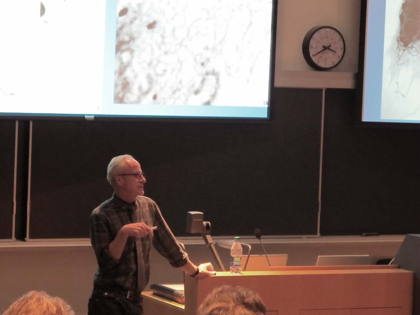 a description of how the development and use of the light and electron microscope has changed our perception of what the inside of a cell looks like. He approached the topic from a historical perspective and summarized the problems encountered by scientists in getting biological tissues into the microscope to view, and how these problems were solved. He finished his presentation by showing examples of how scientists in the LSI are using the electron microscope to visualize the ‘nanospace’ world and how these studies contribute to understanding normal cell function and disease.
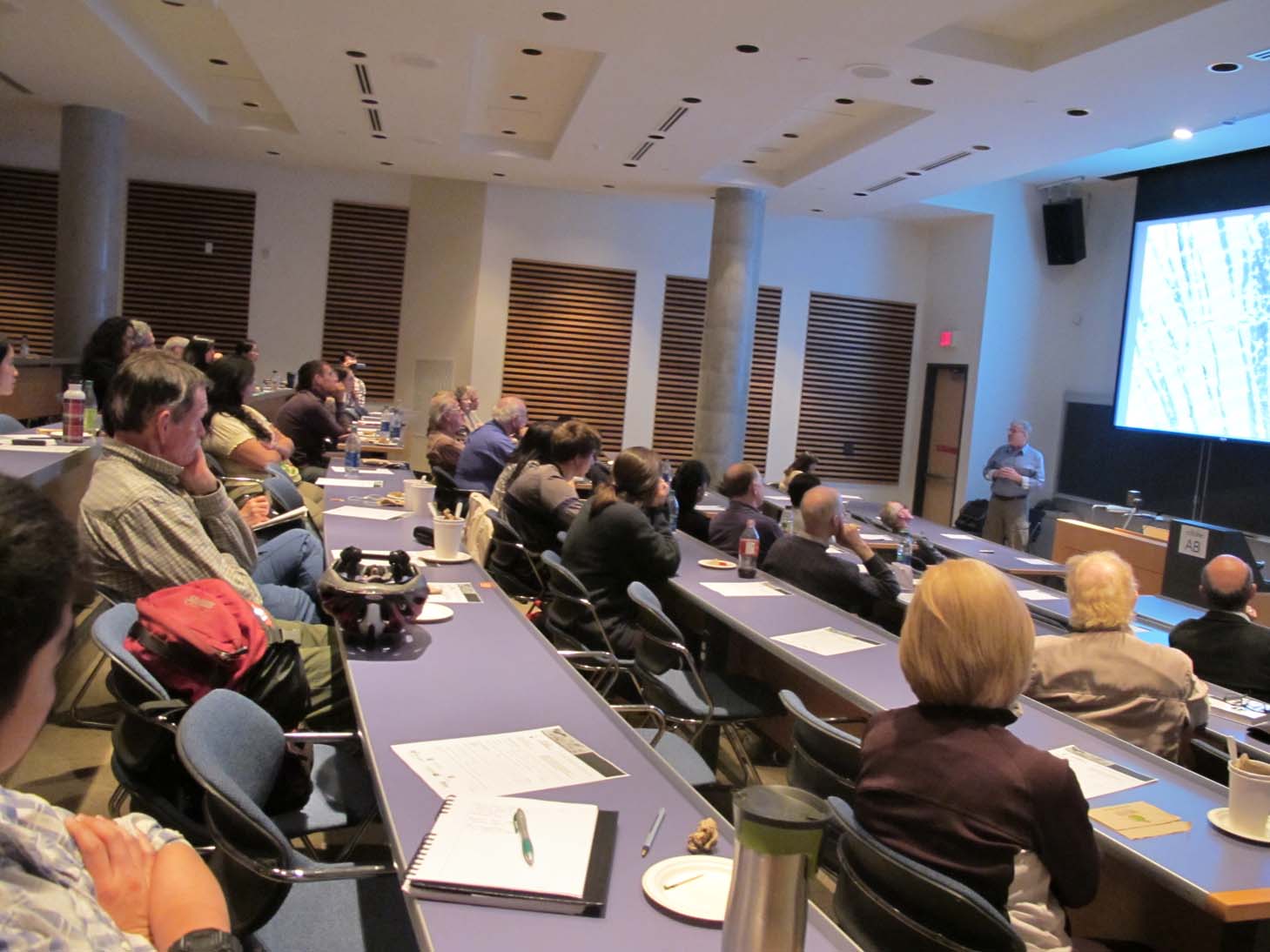 Dr. Edwin Moore discussed nanospaces in heart muscle cells, and how the processes within them control the force with which the heart contracts in a coordinated rhythmic fashion. Dr. Moore explained how molecular organization within nanospaces is key to understanding their function and went on to demonstrate new optical and electron microscopy techniques, and how the images generated are revolutionizing our understanding of cellular structure and function.
A mounted image entitled “The Heart Cell”, supplied by Dr. Moore, was given away as the door 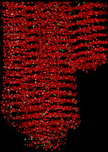 prize to a member of the audience.
The LSI Café Scientifique is co-sponsored by the Life Sciences Institute (LSI), Michael Smith Foundation for Health Research (MSFHR), Faculty of Medicine Research Office, Faculty of Science Dean’s Office, the Leica Corporation, the microscope company Systems for Research and Café Perugia (UBC Food Services).
The next Café Scientific that will continue the “Seeing is Believing” series will take place in the fall of 2012.
To look at more pictures, please visit our facebook page.
“NanoSpace Invaders” by Dr. Wayne Vogl
NanoSpace Invaders Pt. 1 from Life Sciences Institute on Vimeo.
“NanoSpace Invaders” by Dr. Ed Moore
NanoSpace Invaders Part 2 from Life Sciences Institute on Vimeo.
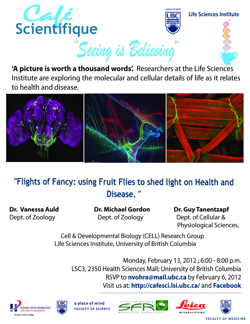
Seeing is Believing Part 2 – “Flights of Fancy: using Fruit Flies to shed light on Health and Disease”
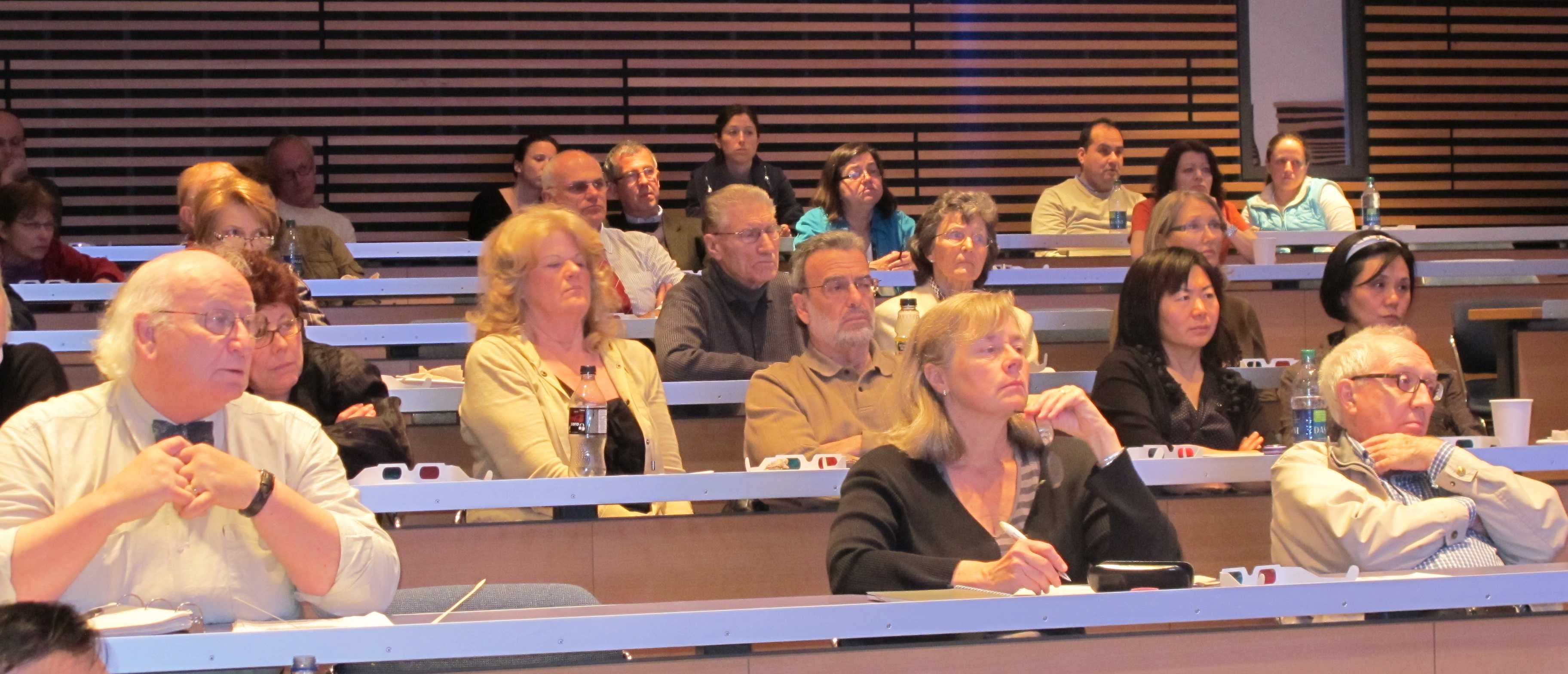
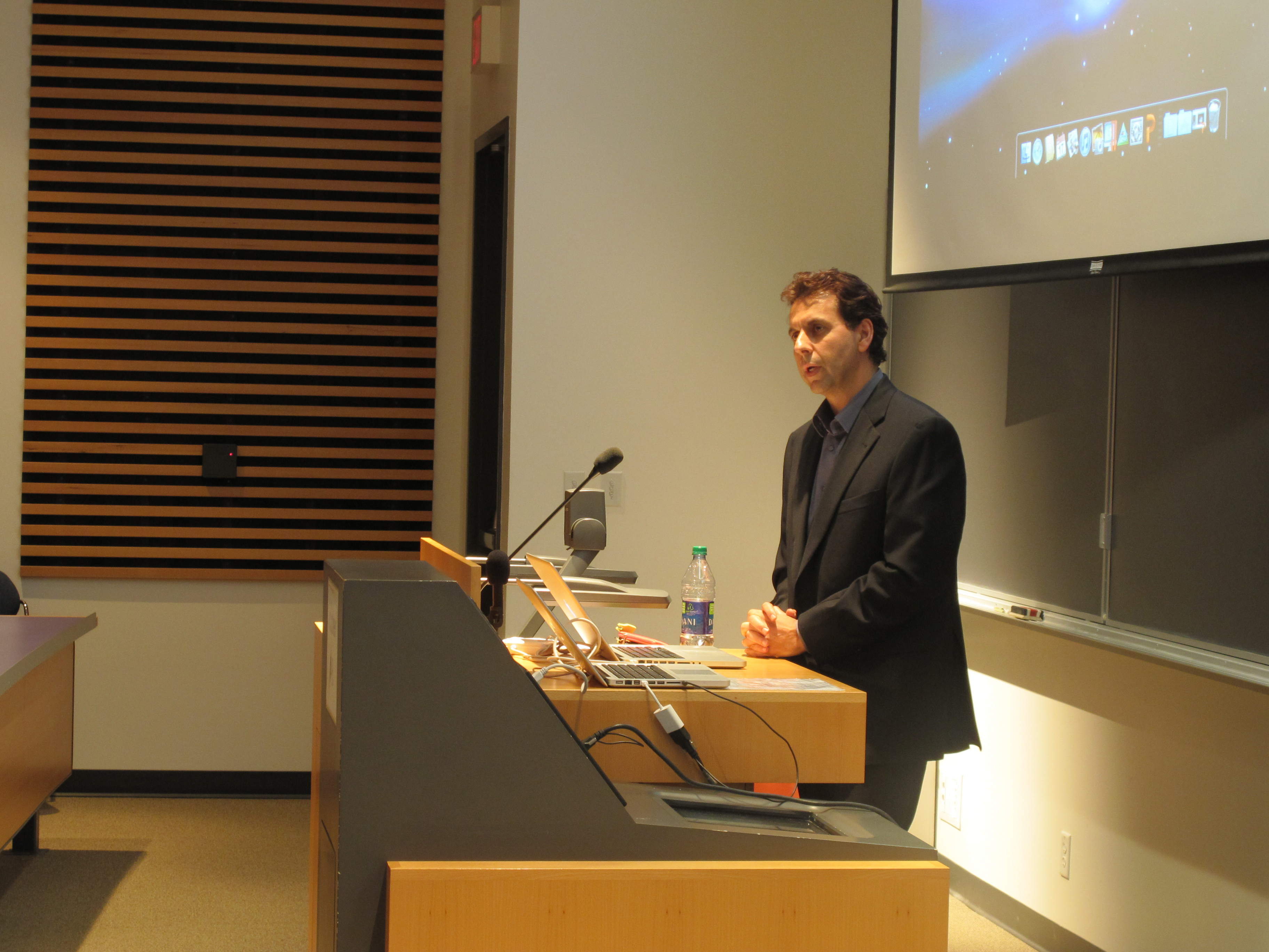
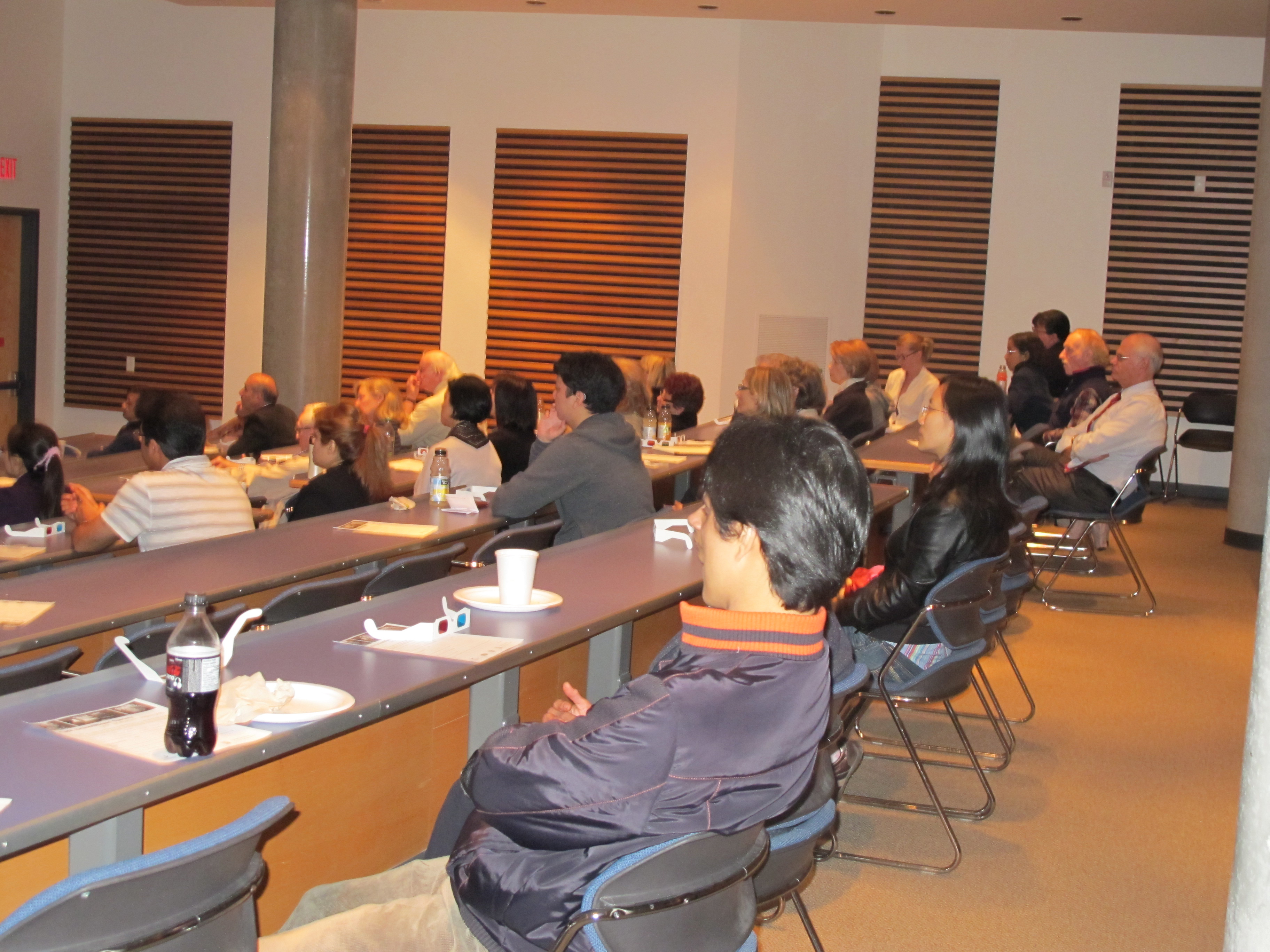
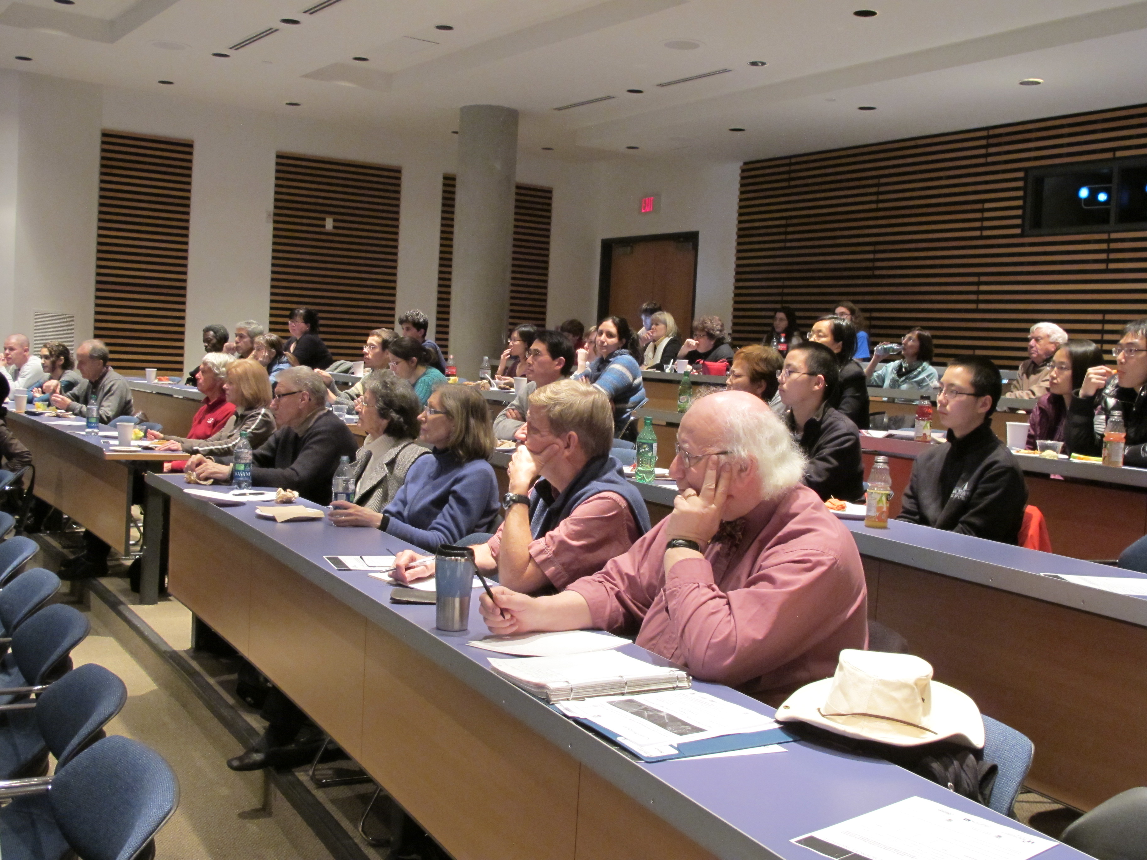 On February 13, 2012, the Life Sciences Institute (LSI) at the University of British Columbia hosted the 9th LSI Café Scientifique. Over 50 interested community members, students and faculty gathered for an informal and participatory dialogue with LSI experts for the second session in the “Seeing Is Believing” series. The particular topic of the session was “Using Fruit Flies to shed light on Health & Disease”.
The Café featured members of the Cell and Developmental Biology Research Group who use Drosophila melanogaster (fruit fly) as their genetic model organism of choice to understand molecular interactions that occur during nervous system and connective tissue development. The interactive session was presented by Drs. Vanessa Auld, and Michael Gordon, from the department of Zoology and Dr. Guy Tanentzapf from the Department of Cellular and Physiological Sciences.
Dr. Vanessa Auld started the presentation with an overview of the Drosophila model system and why it makes an excellent model to study development and disease progression. As case in points is using this system to understanding how the nervous system develops and functions. The Drosophila nervous system is far simpler and with the ease of genetic manipulations in Drosophila this provides a model in which to test the function of genes during development and a model of a range of human diseases. She also introduced the audience to a history of green fluorescent protein and the impact of this protein and its derivatives on modern cell and developmental biology research. The ability of this protein to tag cells and individual proteins allows for imaging the development and disease progression in living animals. Dr. Auld then briefly touched on her research program and why her lab investigates the roles that glia play in the development and function of the nervous system. Dr. Auld explained that glia fulfill a number of important functions including generating an insulating barrier to isolate one neuron from another, to protect neurons from environmental changes and pathogens and to provide structural support. In order to study the molecular and cellular interactions that occur between glia and neurons during development, the Auld lab studies the fruit-fly, Drosophila melanogaster, because there are many parallels between the glia of vertebrates and Drosophila melanogaster.
Dr. Guy Tanentzapf discussed how his lab uses molecular biology, genetics, and cutting edge imaging technology to address basic biological questions. The Tanentzapf lab is mainly interested in the study of cell adhesion, the process by which cells attach to their surrounding environment, in particular how cell adhesion contributes to muscle function. The fruit fly serves a powerful model system for studying integrin function and components of integrin-mediated adhesion are structurally and functionally similar to their counterparts in vertebrates. Compared to vertebrate genomes the fly genome is simpler and there are fewer components of the integrin adhesion complex as well as less redundancy. In addition to providing basic insight into how animals develop and retain their structures, the study of the role of integrins in the fly also provides knowledge that will help improve human health.
Dr. Michael Gordon discussed how the mechanisms brains use to process diverse sensory stimuli, form internal representations of the outside world, and generate appropriate behavioural actions remain some of the great mysteries of biology. Because the difficulty of the problem scales with the complexity of the nervous system, Dr. Gordon has chosen to study it in the fruit fly, which uses only ~100,000 neurons (1 million times fewer than humans) to generate a complex array of behaviours. The fly also offers a wide and ever-growing array of molecular and genetic tools to probe both the neural circuits and molecules underlying sensory processing and behaviour. Dr. Gordon explained how understanding the fly’s brain will give insight into how circuits are organized and function in our own brains, and how evolution has sculpted solutions to common problems like how to locate food, decide what to eat, or find a mate. 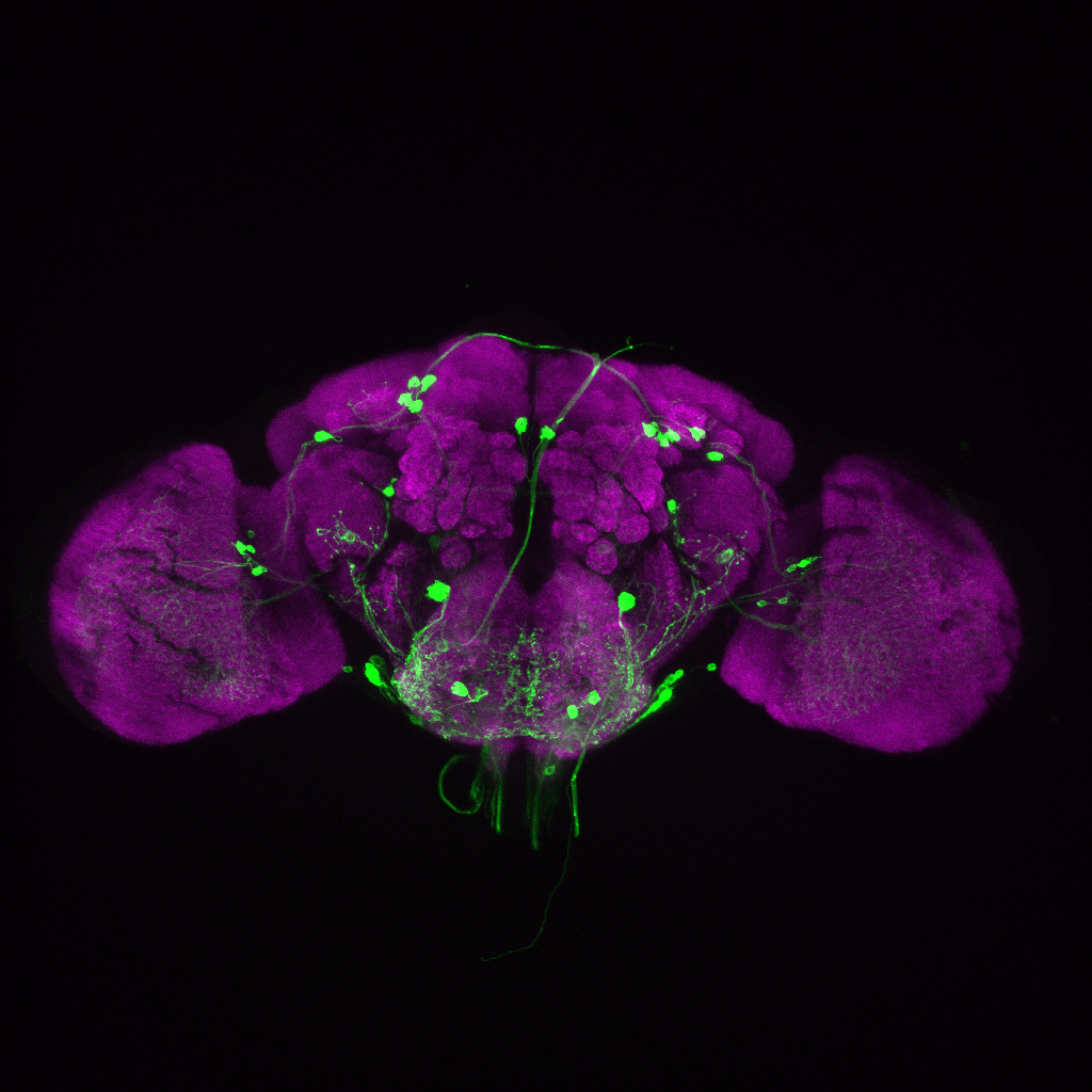
A mounted image entitled “The brain of a fruit fly, Drosophila melanogaster”, supplied by Dr. Gordon, was given away as the door prize to a member of the audience.
The LSI Café Scientifique is co-sponsored by the Life Sciences Institute (LSI), Michael Smith Foundation for Health Research (MSFHR), Faculty of Medicine Research Office, Faculty of Science, the Leica Corporation, the microscope company Systems for Research and Café Perugia (UBC Food Services).
The next Café Scientific that will continue the “Seeing is Believing” series will take place in spring 2012.
To view more pictures from the event, please visit our facebook page.
“Using fruitflies to visualize development” by Dr. Vanessa Auld
“Visualizing how Muscles Connect to Tendons in the Fly” – Dr. Guy Tanentzapf
“Looking into the mind of the Fly” by Dr. Mike Gordon
Seeing is Believing Part 1, The Universe Within – Seeing how cells move and interact.
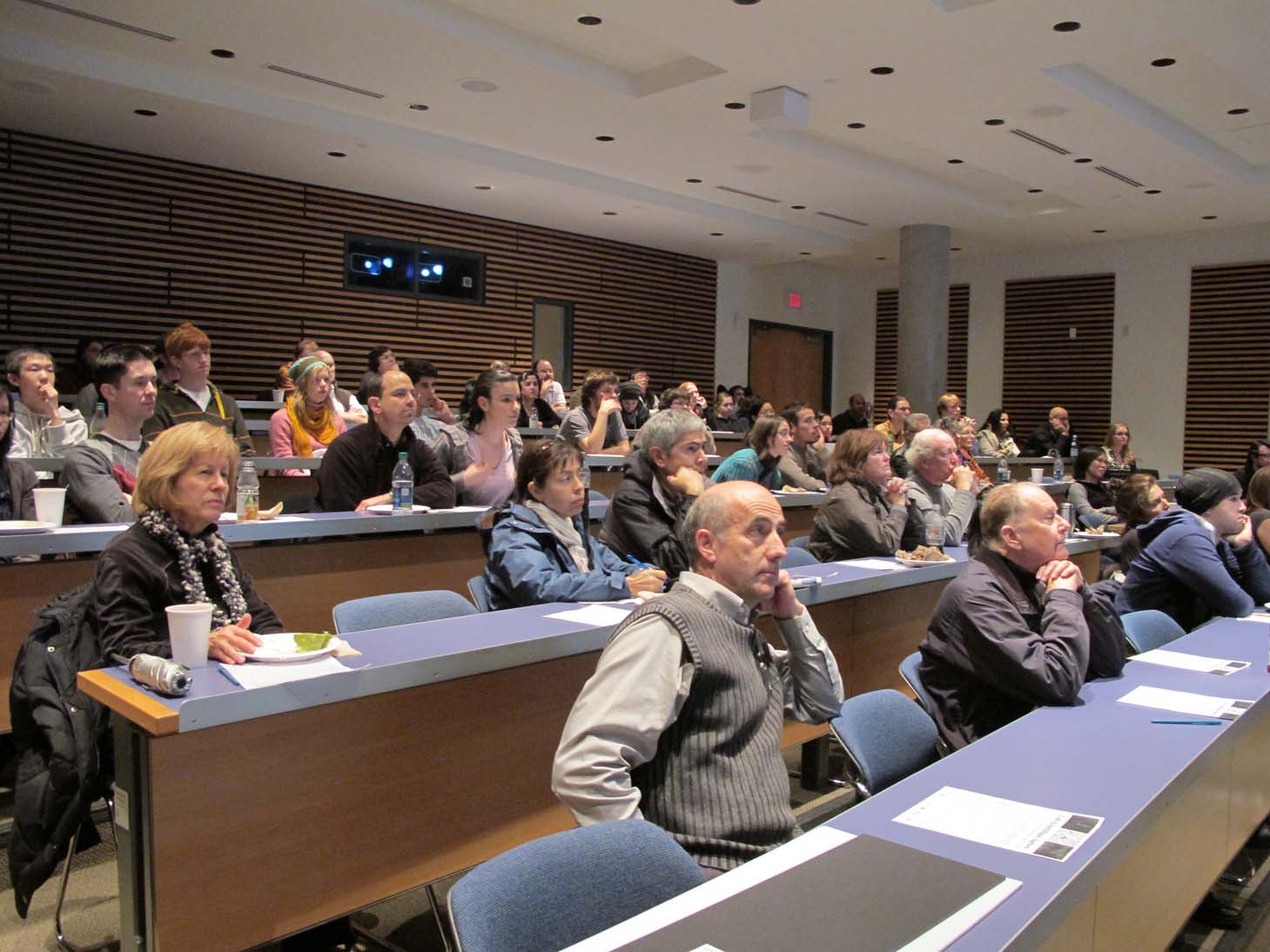 On November 21, 2011, the Life Sciences Institute (LSI) at the University of British Columbia hosted the 8th LSI Café Scientifique. A record number of over 60 interested community members, students and faculty gathered for an informal and participatory dialogue with LSI experts for the first session of the “Seeing Is Believing” series. The particular topic of the session was “Imaging the Developing and Aging Brain”.
The Café featured members of the Cell and Developmental Biology Research Group and the Brain Research Centre who discussed state-of-the-art imaging and genetic technologies and how these are being used to understand how brain networks form and function. The interactive session was presented by Drs. Kurt Haas, Shernaz Bamji and Douglas Allan, all from the Department of Cellular and Physiological Sciences.
Dr. Kurt Haas started the presentation with a brief introduction to neuroscience research and how innovations in imaging technologies have enabled us to peer into the living brain. He discussed his own research using direct imaging of the growth of neurons in the developing brain of tadpoles. Dr. Haas showed movies of these cells dynamically extending and retracting branches as they seek other cells to form neural circuits. He discussed the complexities of this process and how errors in brain cell growth and connectivity can lead to common neurodevelopmental disorders like Autism, Schizophrenia, and Epilepsy.
Dr. Shernaz Bamji discussed how nerve cells communicate at specialized junctions called 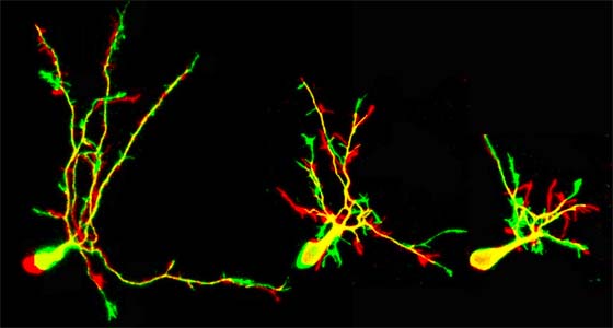 “synapses” and how advances in imaging and cell culture technologies have enabled researchers to track the movement of proteins and to watch as synapses form and function. She further discussed how altering the state of adhesion at synapses could lead to abnormalities in learning and memory that may underlie disorders such as Huntington’s and Alzheimer’s diseases.
Dr. Douglas Allan gave an overview of recent advances in molecular genetic technologies that are used to image and manipulate gene expression in select populations of neurons in the brain. He discussed the techniques of transgenesis, and showed how DNA that had previously been known as junk DNA is in fact used to regulate gene expression and can be used to express fluorescent proteins in specific types of neurons. Finally, he discussed the use of multiple fluorescent proteins and DNA-manipulating enzymes to generate multi-coloured neuronal tissues that can be used to map brain circuits.
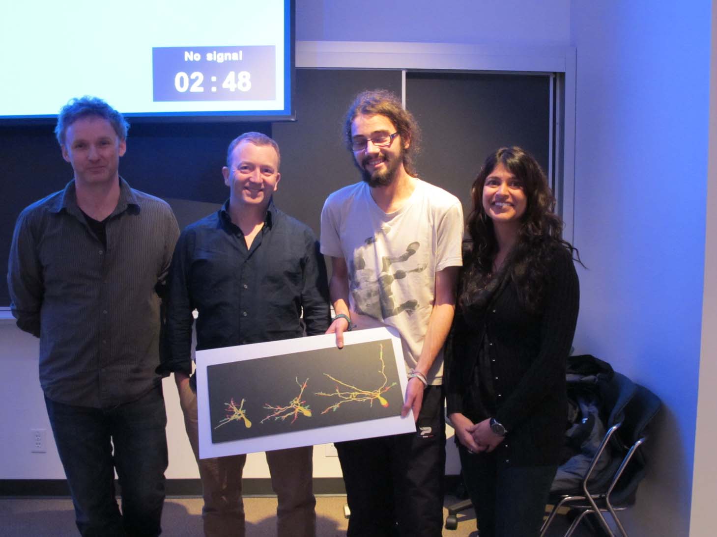
A mounted image entitled “Growth of a Brain Neuron”, supplied by Dr. Haas, was given away as the door prize to a member of the audience.
 The LSI Café Scientifique is co-sponsored by the Life Sciences Institute (LSI), Michael Smith Foundation for Health Research (MSFHR), Faculty of Medicine Research Office, Faculty of Science, Leica Microsystems, Systems for Research and Café Perugia (UBC Food Services).
The next Café Scientific that will continue the “Seeing is Believing” series will take place in early 2012.
To view more pictures from the event, please visit our facebook page.
Seeing is Believing by Dr. Kurt Haas
Seeing is believing by Dr. Shernaz Bamji
Seeing is believing by Dr. Doug Allan
E-natomy – How digital anatomy has changed the way we teach and practice medicine.
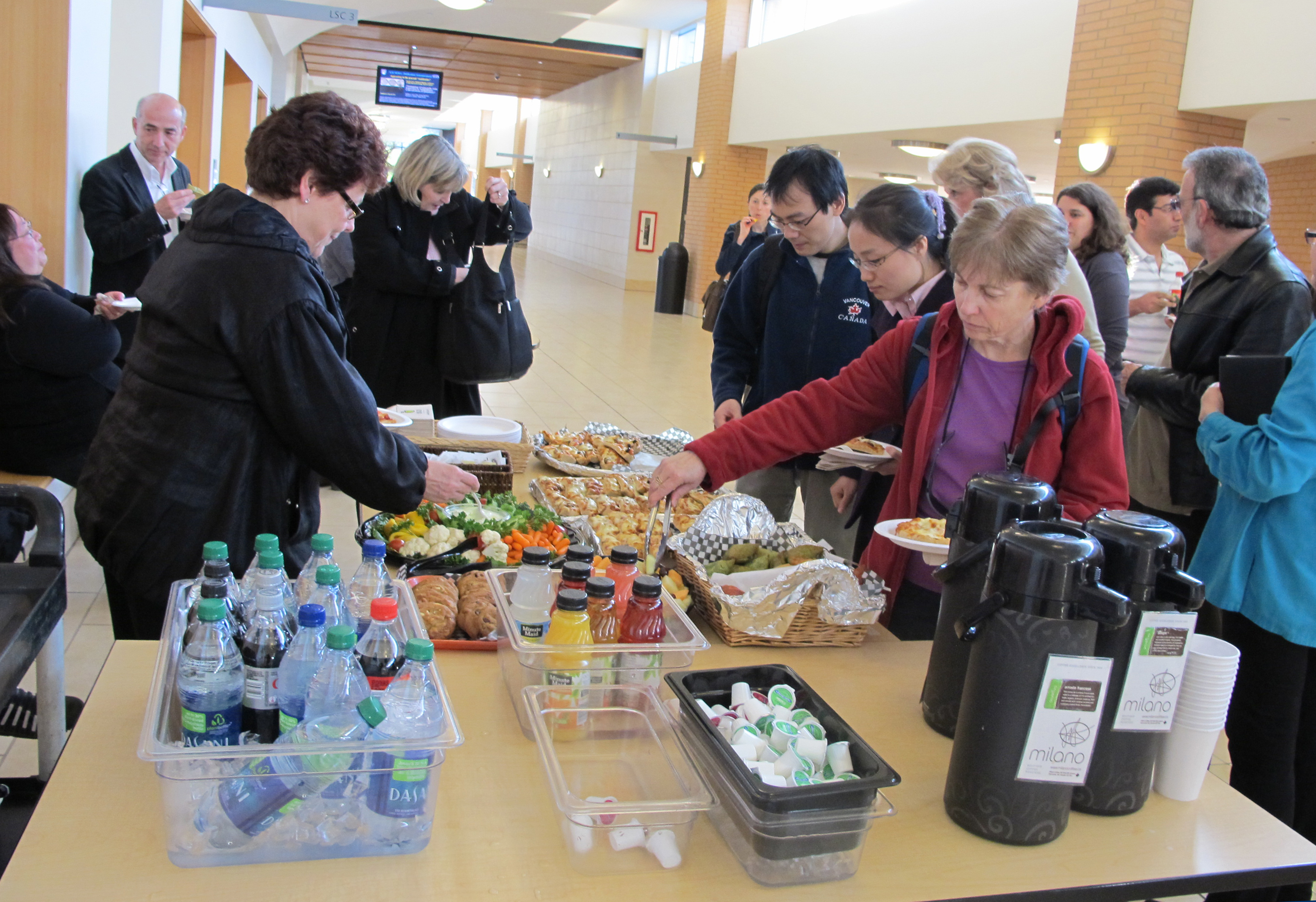
 On May 17, 2011, the Life Sciences Institute (LSI) at the University of British Columbia hosted the seventh LSI Café Scientifique. Over 50 interested community members, as well as students and faculty gathered for an informal and participatory dialogue with experts on the topic of “E-natomy – How digital anatomy has changed the way we teach and practice medicine?”. The Cafes are sponsored by the LSI, The Michael Smith Foundation for Health Research, the Faculties of Medicine and Science and Café Perugia (UBC Food Services).
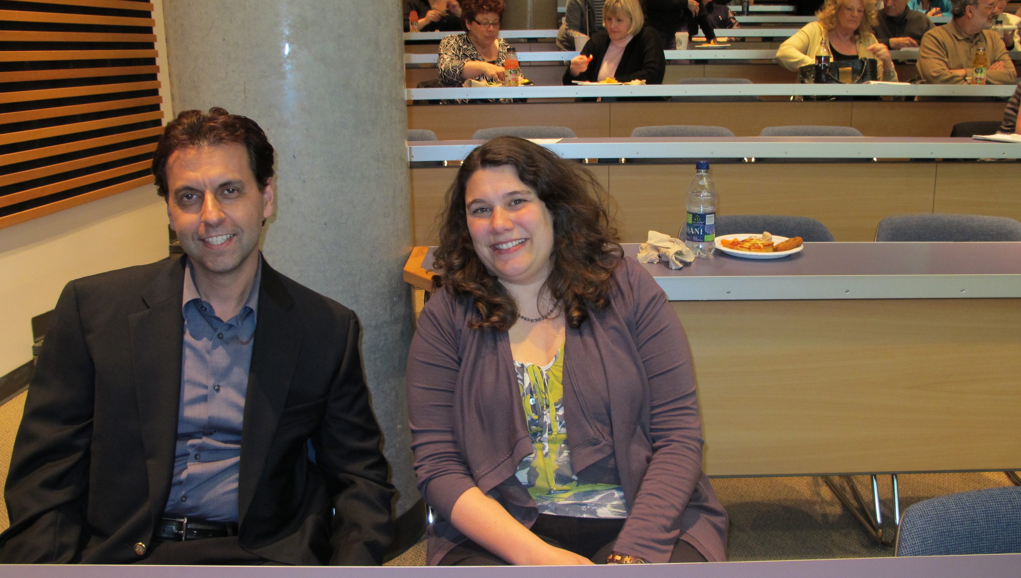
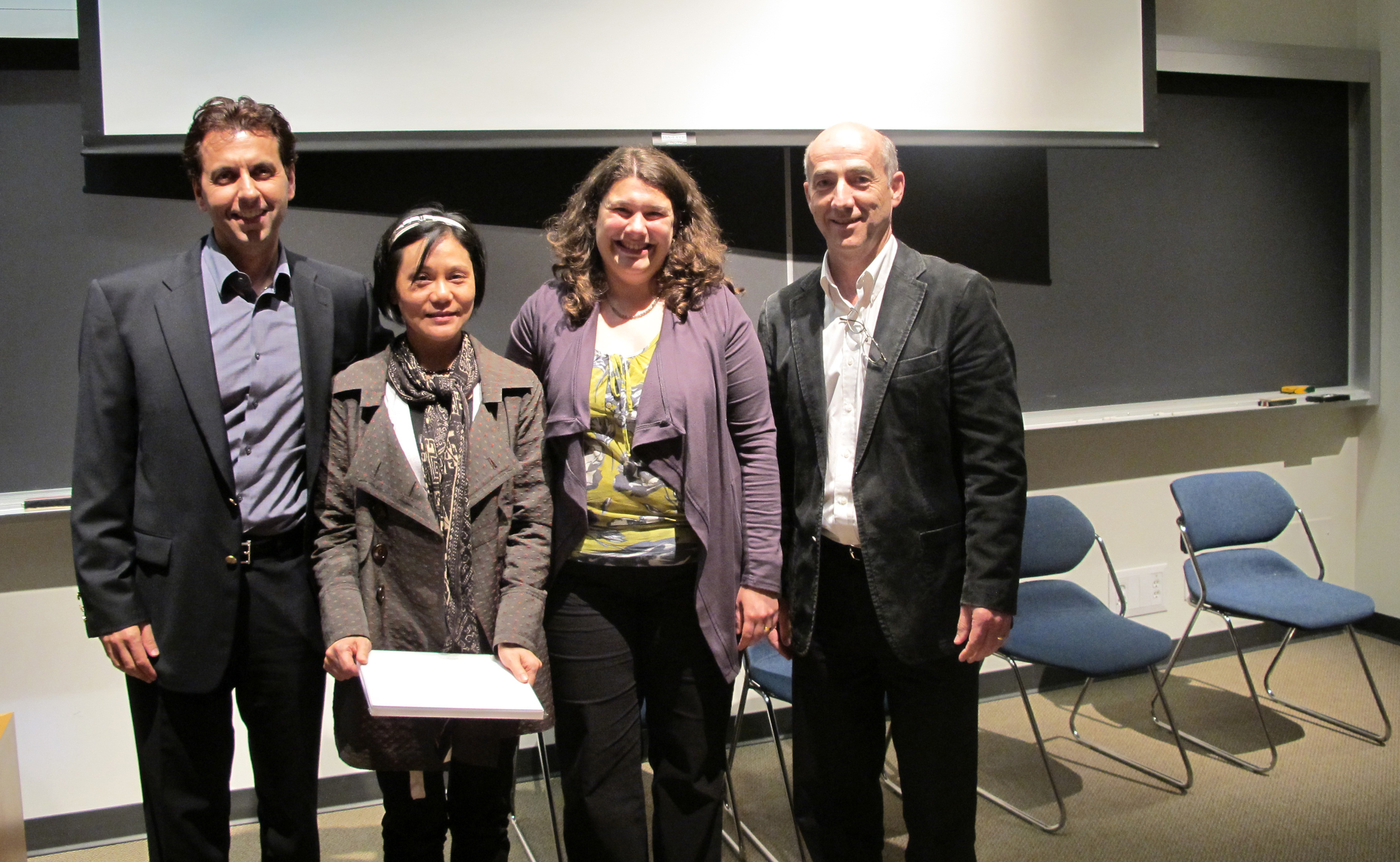
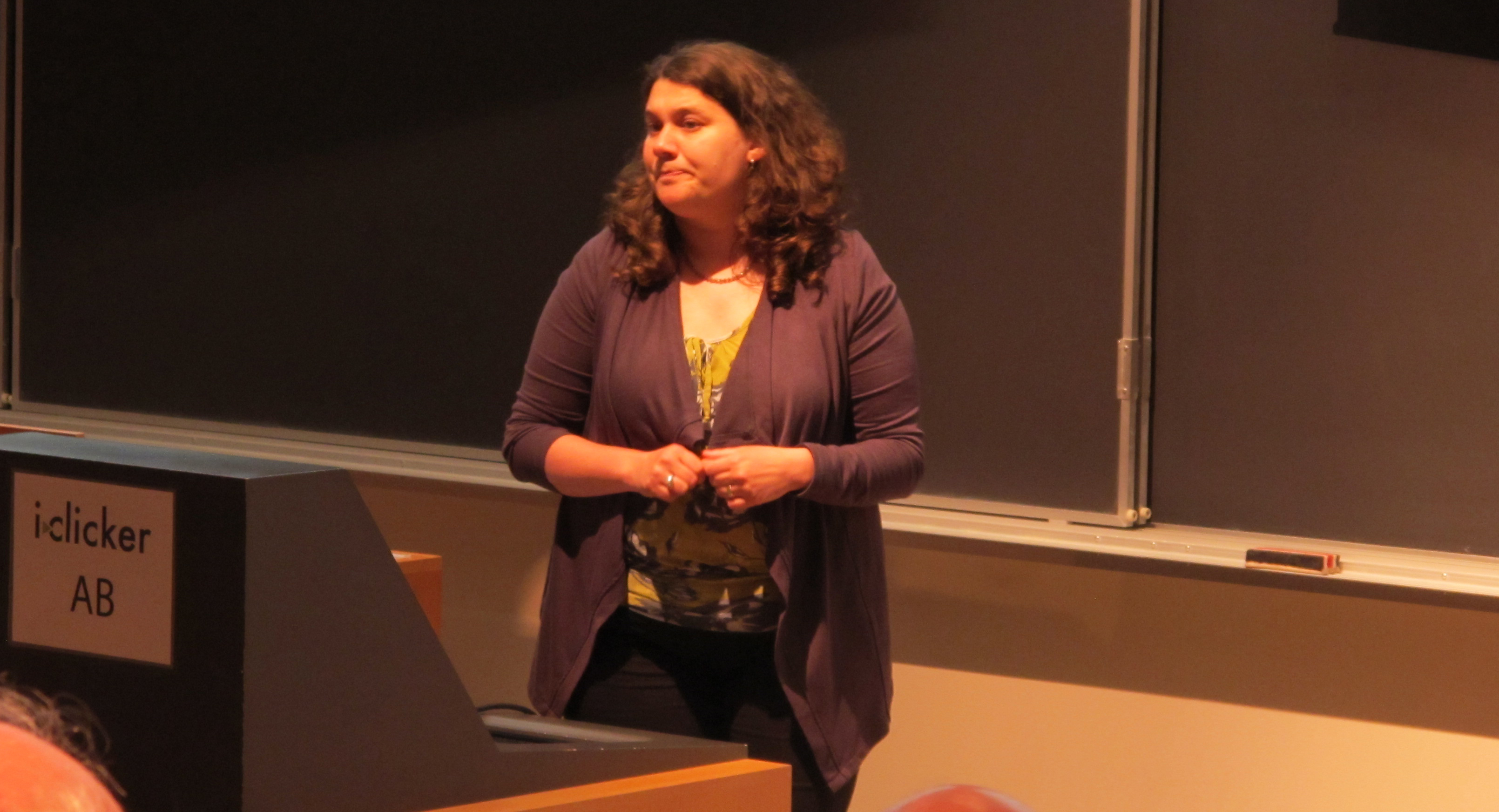
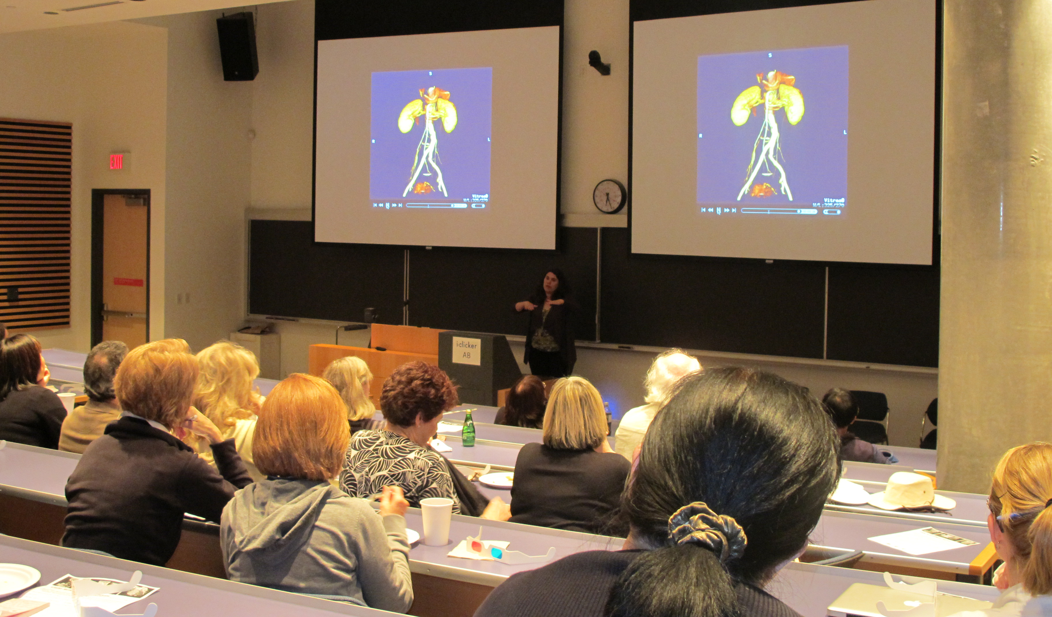
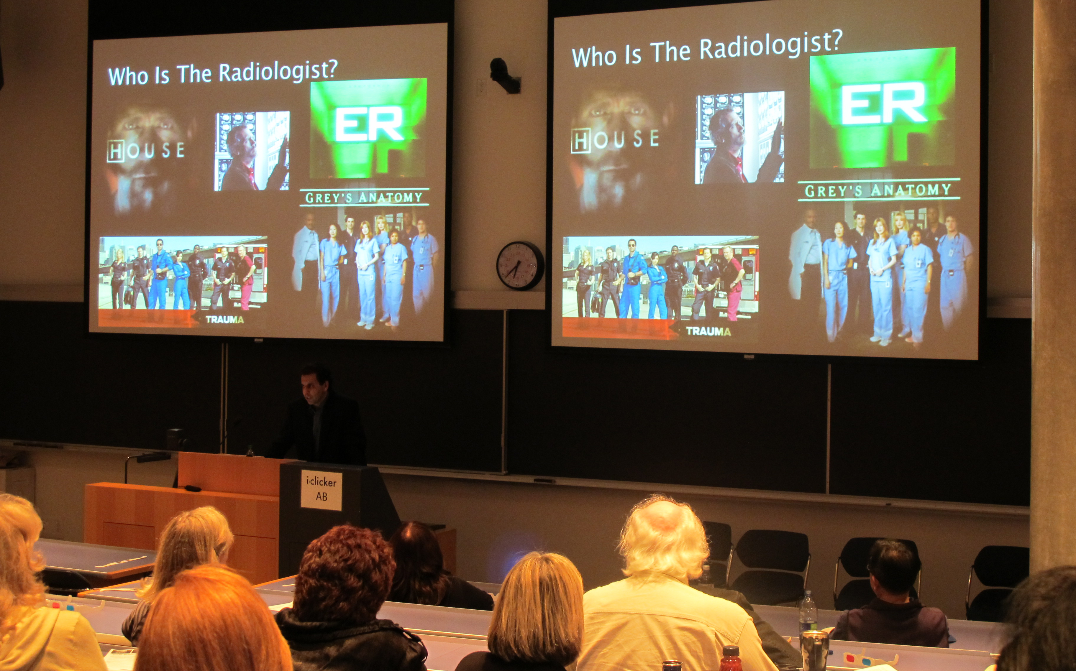
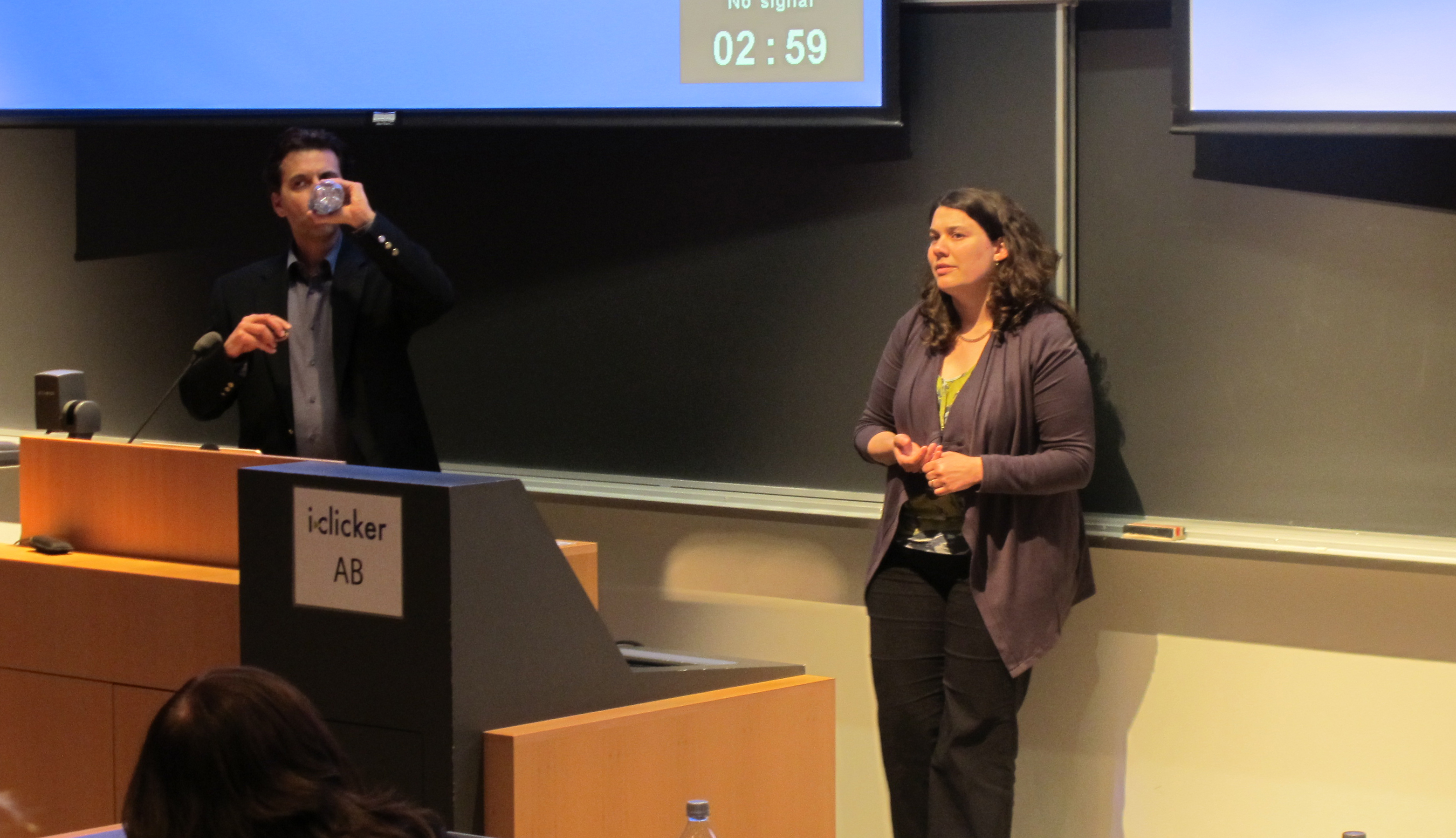
This Café featured two members of the Faculty of Medicine who use digital anatomy in teaching medical students and in clinical practice. This interactive session was presented by Dr. Claudia Krebs, a Senior Instructor in the Department of Cellular & Physiological Sciences, and Dr. Savvas Nicolaou, Associate Professor and Director of Emergency Radiology, practicing at the Vancouver General Hospital. Drs. Krebs and Nicolaou each explained how the advances in whole body imaging have changed the way students learn anatomy and how patients are treated in the ER. Real-life examples of digital anatomy’s role in teaching as well as in practice were shown and the power of CT scans, MRIs and ultrasound scans compared with traditional x-rays was explained.
 New techniques in medical imaging have made it possible to look inside the human body with great precision and with less invasiveness in order to diagnose pathologies that in the past could only be seen with surgical exploration. Learning human anatomy using cadaveric dissection supplemented with x-ray, ultrasound, CT and MRI images allows the physicians of tomorrow to have a better understanding of the 3-dimensional organization of the human musculoskeletal and circulatory systems, internal organs and nervous system. Teaching these new skills in medical school is required to train the modern physician to effectively utilize imaging modalities and interpret pathologies in the clinic today.
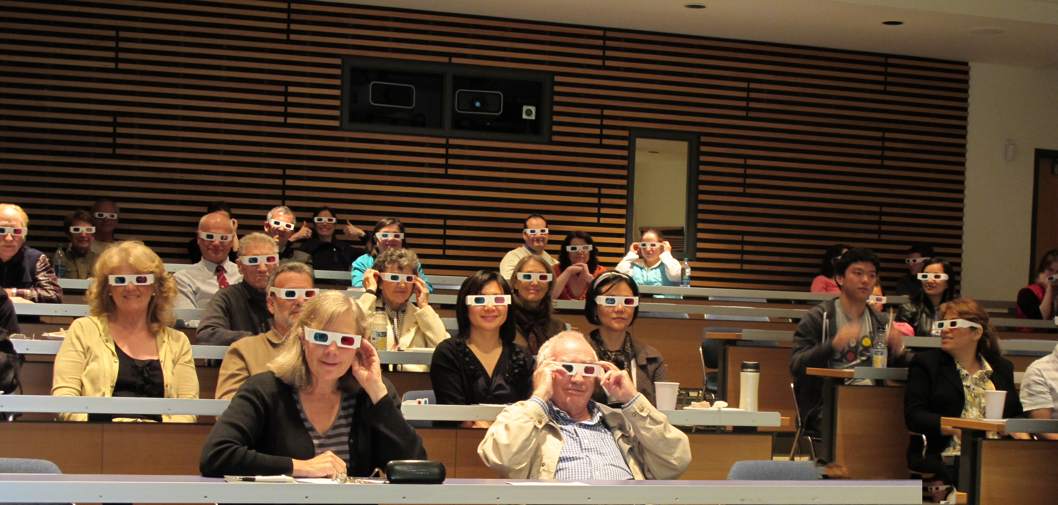
The session was highly interactive and visually entertaining; the presentations included 3D images of the human pelvis, rotating in space which the audience was able to view with 3D glasses. The audience also viewed several staged videos where the power of CT scanning in the Emergency room could be seen as an important diagnostic tool to guide the ER staff in the timely treatment of patients, and in saving lives.
 The door prize awarded to a member of the audience was a copy of the photobook created by Dr. Claudia Krebs with the assistance of Monika Fejtek. This photobook contained digitized images of the original anatomical artwork of the artist Nan Cheney. This collection of pen and ink drawings is an asset of the original Dept of Anatomy at UBC (now called the Dept of Cellular and Physiological Sciences).
Nan Gertrude Lawson Cheney (1897-1985), a contemporary of Emily Carr, was born in Windsor, Nova Scotia and moved to Vancouver in 1937 where she became a well-known portrait and landscape painter. Nan Cheney was also a medical illustrator in the Department of Anatomy at UBC from 1951 to 1956, and continued to work as a medical illustrator until her retirement in 1962. The Department of Cellular and Physiological Sciences currently owns over fifty original works of art by Nan Cheney some of which are exhibited in four glass cases in the main colonnade of the Life Sciences Centre. Each of the works was created from an anatomical dissection done by a staff member in the Department of Anatomy during that time. The Nan Cheney photobook was created to highlight the collection that is displayed in the Colonnade.
Memories in your Genes?
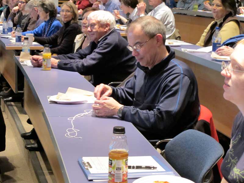 On March 29, 2011, the Life Sciences Institute (LSI) at the University of British Columbia hosted the sixth LSI Café Scientifique. A record number of over 75 interested community members, students and faculty gathered for an informal and participatory dialogue with LSI experts on the topic of “Memories in your Genes?”
The Café featured members of the Molecular Epigenetics Research Group who are studying epigenetic gene regulation. The interactive session was presented by Dr. Carolyn Brown, Professor and Head of the Department of Medical Genetics; Dr. Louis Lefebvre, Assistant Professor in Department of Medical Genetics; and Dr. LeAnn Howe, Associate Professor in Department of Biochemistry and Molecular Biology.
There are the some 3 billion basepairs of DNA in each cell of your body, yet cells remember how they have been instructed to act – make an eye or make a kidney. Epigenetics is the study of the identifying marks that are placed on the packaged DNA that are passed on through cell divisions – and sometimes even through generations.
Dr. Brown started the presentation with a brief introduction of terminology and gave an overview of how genes inherit instructions. Drs. Howe, Lefebvre and Brown then presented their epigenetic research stories.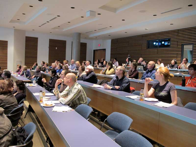
Dr. Howe discussed how HATs (histone acetyltransferases) make the genes stand out in a crowd and how histone acetylation is preserved through DNA replication. Dr. Lefebvre gave an overview of maternal and paternal genes, how the genetic information is written in the DNA molecule, the characteristic number of chromosomes in the cells, maintenance of methylation marks at replication and how some genes become imprinted during evolution. Dr. Brown gave answers to why calico cats are almost always female and demonstrated how the calico cat gets its spots.
A mounted image entitled “Activity of an imprinted gene in a germ cell, the oocyte”, supplied by Dr. Lefebvre, was given away as the door prize to one of the member of the audience.
The LSI Café Scientifique is co-sponsored by the Life Sciences Institute (LSI), Michael Smith Foundation for Health Research (MSFHR), Faculty of Medicine Research Office, Faculty of Science and Café Perugia (UBC Food Services).
The next Café Scientific is entitled “E-natomy – How digital anatomy has changed the way we teach and practice medicine” and will take place on May 17th at 6PM. Other future topics include discussions on how streamlining drug development is important, the innovations in safe blood products and blood substitutes, and brain development.
Introduction by Dr. Carolyn Brown
“How HATs make your genes stand out in crowd”
by Dr. LeAnn Howe
“How mom and dad contribute differently to your epigenome”
by Dr. Louis Lefebvre
“Why calico cats are always female”
by Dr. Carolyn Brown