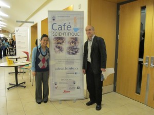
 On Nov 14, 2012, the Life Sciences Institute (LSI) at the University of British Columbia hosted the 11th LSI Café Scientifique. Over 70 interested community members, students and faculty gathered for an informal and participatory dialogue with LSI experts for the fourth session in the “Seeing Is Believing” series. The topic of the session was “Viewing the Biological World with X-rays and Magnetic Fields”.
The Café featured Dr. Michael Murphy, a member of the Bacterial Adaptation & Response Networks (BARN) LSI Research Group and the Dept of Microbiology & Immunology, and Dr. Lawrence McIntosh, a member of the Chemical Biology of Disease (CBD) LSI Research Group and the Depts of Biochemistry & Molecular Biology and Chemistry.
Dr. Michael Murphy started the discussion explaining the ranges of sizes of objects, starting with a grain of rice and moving progressively smaller and smaller to show where biomolecules fit into the sequence of sizes. He then went on to show that X-ray crystallography provides a detailed atomic view of biological molecules. This visualization technique relies on producing small crystals of the biomolecule that are subject to an intense highly focused X-ray beam. The data collected from the X-ray beam interacting with the crystal is used to create three-dimensional maps of the electron density that describe the structure of the molecules. The resulting image is a snapshot of an average over all the molecules in the crystal. Multiple snapshots of the crystallized biomolecules in different states such as a free receptor versus the ligand bound form can be used to describe function of the proteins at a molecular level. Examples were taken from one of the systems studied in the Murphy lab, the human bacterial pathogen, multiple drug resistant Staphylococcus aureus. Bacteria need to acquire iron to grow but our bodies attempt to sequester iron to limit bacterial growth. Effective pathogens like S. aureus have developed specialized scavenger proteins to overcome this nutritional need by pirating iron from host proteins. These bacterial scavenger proteins are potential targets for the development of new antibacterial therapeutics and diagnostic drugs. Throughout the talk, computer generated protein structures were shown that highlighted the various regions of proteins using a variety of views. The talk ended with a brief question and answer session.
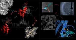 Dr. Lawrence McIntosh went on to explain that Nuclear Magnetic Resonance (NMR) spectroscopy is a “molecular microscope” that enables researchers to study the three-dimensional structures of proteins and thereby gain insights into their biological functions. NMR relies on measuring the energy required to “flip” the spins of nuclei aligned within a powerful magnet field. This energy depends on the chemical environment of the nucleus, and hence on the structure of protein. Importantly, proteins do not have static structures, as shown in the field by beautiful diagrams of ribbons and coils, but rather are highly flexible and dynamic. This flexibility, which can also be measured with NMR, is important for protein function. One such function is the self-inhibition of a transcription factor that balances the energetic cost of unfolding a helix structure of this protein with the benefit of binding DNA. This balance can be changed in response to cellular signals in order to turn genes on or off, working as a molecular switch. Dr. McIntosh’s talk also highlighted computer generated molecular models that showed the dynamic structure of the proteins he studies. At the end, he also answered questions from the audience.
 Both the Murphy lab and the McIntosh lab are aided by infrastructure (equipment) purchased by the Canada Foundation for Innovation grant to the ASTRID (“Advanced Structural Analysis of Re-emerging Infectious Diseases”) initiative, located in the Life Science Centre.
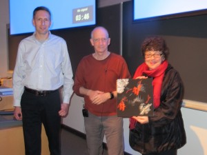
At the end of the talks, a mounted image entitled “Biological NMR Spectroscopy”, supplied by Dr. Lawrence McIntosh, was given away as the door prize to a member of the audience.
The LSI Café Scientifique is co-sponsored by the Life Sciences Institute (LSI), Michael Smith Foundation for Health Research (MSFHR), Faculty of Medicine Research Office, Faculty of Science Dean’s Office, the Leica Corporation, the microscope company Systems for Research and Café Perugia (UBC Food Services).
To look at more pictures, please visit our facebook page.
The next Café Scientific that will continue the “Seeing is Believing” series will take place in early 2013.
“Viewing the Biological World with X-rays and Magnetic Fields” by Dr. Michael Murphy
“Viewing the Biological World with X-rays and Magnetic Fields” by Dr. Lawrence McIntosh

